

Introduction: Understanding the Cell Cycle and CFSE T Cell Proliferation Assay
The cell cycle and CFSE T cell proliferation assay are central subjects in fashionable biology, particularly in cell biology, immunology, and biomedical analysis. Each residing organism will depend on managed cell division to develop, restore tissues, and reply to environmental adjustments.
The cell cycle refers back to the ordered sequence of occasions {that a} cell undergoes to duplicate its DNA and divide into two daughter cells. This course of happens all through an organism’s life and is fastidiously regulated to take care of mobile well being and genetic stability.
Cell division is important for:
Embryonic improvement from a single fertilized egg (zygote)
Development of tissues and organs
Substitute of broken or useless cells
Immune system activation
Upkeep of organ operate
Therapeutic of wounds and accidents
In scientific analysis, understanding how cells divide is extraordinarily essential. One highly effective experimental methodology used to measure immune cell division is the CFSE T cell proliferation assay, which makes use of fluorescent labeling mixed with circulate cytometry.
This system permits scientists to:
Monitor what number of instances cells divide
Measure immune cell activation
Research illness development
Consider responses to remedies
The cell cycle is a extremely managed organic course of consisting of a number of sequential phases. Every part ensures that the cell is absolutely ready earlier than progressing to the following stage.
Cells could both stay inactive or actively divide relying on the physiological wants of the organism.
1. Hole 0 (G0) Section – The Resting State
The G0 part is taken into account a non-dividing stage of the cell cycle.
Essential options of the G0 part embody:
Cells are metabolically energetic however not dividing
Cells carry out specialised features
Many differentiated cells stay in G0 completely
Some cells can re-enter the cycle when stimulated
Examples of cells typically in G0:
This stage helps the physique preserve vitality and keep mobile stability.
The G1 part is the primary stage of energetic cell cycle development.
Throughout G1, a number of vital processes happen:
The cell will increase in measurement
Proteins required for DNA replication are synthesized
Mobile organelles multiply
The cell evaluates environmental situations
DNA integrity is checked
Key checkpoint occasions embody:
If situations are unfavorable, the cell could return to the G0 part as an alternative of constant the cycle.
3. S Section – DNA Synthesis
The S part is when probably the most vital occasion of the cell cycle happens: DNA replication.
Main occasions in the course of the S part embody:
Duplication of chromosomes
Formation of sister chromatids
Replication of histone proteins
Elevated DNA content material contained in the nucleus
Preparation for correct cell division
On the finish of this part, every chromosome consists of two similar chromatids.
4. Hole 2 (G2) Section – Last Preparations for Division
The G2 part ensures that the cell is totally prepared for mitosis.
Essential processes embody:
Verification of profitable DNA replication
Restore of DNA injury
Synthesis of mitotic proteins
Group of microtubules
Preparation of spindle equipment
This checkpoint prevents broken or incomplete DNA from being handed to daughter cells.
5. M Section – Mitosis and Cytokinesis
The M part represents the precise division of the cell.
This part consists of two main processes:
Mitosis
Mitosis contains a number of phases:
Prophase
Metaphase
Anaphase
Telophase
Throughout mitosis:
Chromosomes condense
Nuclear envelope breaks down
Chromosomes align on the middle
Sister chromatids separate
Cytokinesis
Cytokinesis is the ultimate step the place:
Understanding the cell cycle and CFSE T cell proliferation assay is extraordinarily essential in a number of scientific disciplines.
In Immunology
Cell cycle evaluation helps researchers perceive immune responses corresponding to:
Activation of T cells throughout an infection
Enlargement of immune cell populations
Immune response to vaccines
Detection of immune problems
T cells proliferate after they acknowledge pathogens or irregular cells.
In Most cancers Biology
Most cancers is primarily brought on by uncontrolled cell division.
Cell cycle evaluation helps scientists:
Research tumor progress
Establish irregular proliferation patterns
Consider most cancers remedies
Develop focused therapies
In Medical Analysis
Researchers use cell proliferation assays to:
Research autoimmune ailments
Examine inflammatory responses
Consider drug results
Develop new immunotherapies
The CFSE T cell proliferation assay is a extensively used method for monitoring cell division in immunology experiments.
CFSE stands for:
Carboxyfluorescein Succinimidyl Ester
This fluorescent dye permits scientists to look at how cells divide over time.
How CFSE Works
The mechanism of CFSE staining includes a number of essential steps:
CFSE enters dwell cells simply.
The dye binds completely to intracellular proteins.
All cells initially show the identical fluorescence depth.
When a cell divides:
Every new technology has half the fluorescence of the earlier one.
Consequently:
First technology cells present highest fluorescence
Second technology reveals decreased fluorescence
Later generations present progressively decrease fluorescence
This sample kinds a number of peaks in circulate cytometry evaluation.
Movement cytometry is a vital analytical method used within the cell cycle and CFSE T cell proliferation assay.
This expertise permits fast evaluation of hundreds of cells individually.
What Movement Cytometry Measures
Movement cytometry can measure:
Cell measurement
Cell complexity
Fluorescence depth
Floor protein markers
Intracellular proteins
Essential Movement Cytometry Parameters
Ahead Scatter (FSC)
Signifies cell measurement.
Aspect Scatter (SSC)
Signifies inside complexity.
Fluorescence Channels
Used to detect:
CFSE sign
CD markers
Different fluorescent labels
1. Preparation Stage
Essential preparation steps embody:
Sporting laboratory gloves and protecting clothes
Sterilizing dissection devices
Getting ready HBSS with fetal calf serum
Labeling experimental tubes
Correct preparation ensures experimental accuracy and prevents contamination.
2. Mouse Spleen Dissection
Steps concerned:
Euthanize mouse utilizing carbon dioxide system
Safe mouse on dissection board
Carry out belly incision
Find spleen hooked up to abdomen
Rigorously isolate spleen tissue
Switch spleen into HBSS answer
The spleen is wealthy in immune cells, making it best for proliferation research.
3. Immune Cell Isolation
The aim of this step is to acquire splenocytes.
Process contains:
Putting spleen on cell strainer
Crushing tissue to launch cells
Amassing dissociated cells
Centrifuging to pellet cells
Lysing pink blood cells
Washing immune cells
Adjusting last cell focus
Cells are then prepared for staining and stimulation.
Key steps:
Divide cells into experimental teams
Add CFSE dye
Incubate cells
Wash extra dye
Stimulate chosen samples with anti-CD3 antibody
Tradition cells in incubator
Two teams are analyzed:
Management group:
Stimulated group:
Activation of T cells
Elevated cell division
College students typically battle with circulate cytometry evaluation, so here’s a simplified breakdown.
Step 1: Establish Lymphocytes
Use FSC vs SSC plots.
Step 2: Gate T Cells
Choose CD3 constructive cells.
Step 3: Establish Subsets
Separate:
Step 4: Analyze CFSE Sign
Have a look at histogram peaks representing cell generations.
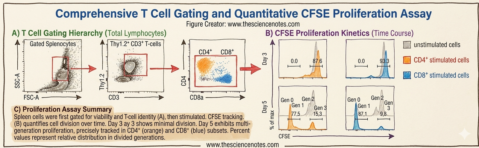
Observations After 3 Days
Observations After 5 Days
Key Experimental Findings
CD4 T cells proliferate after stimulation
CD8 T cells present stronger proliferation
Stimulation considerably will increase cell division
This assay is extensively utilized in fashionable analysis.
Main functions embody:
Different strategies embody:
BrdU Assay
Labels newly synthesized DNA.
EdU Assay
Extra delicate and quicker than BrdU.
Fucci Mouse Mannequin
Permits real-time visualization of cell cycle phases.
Understanding the cell cycle and CFSE T cell proliferation assay is important for college kids finding out life sciences.
Key studying factors embody:
Cell cycle phases regulate cell division
CFSE staining tracks cell proliferation
Movement cytometry gives quantitative evaluation
Immune cells divide in response to stimulation
These strategies are extensively utilized in biomedical analysis